
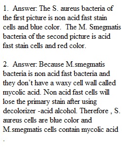
Q This is a group activity. Only one submission per group. BE SURE TO INCLUDE ONLY THE NAMES OF GROUP MEMBERS WHO PARTICIPATED. Answer the following questions using the pictures above. 1. Give the Acid-fast results for each of the two pictures. 2. Why are the S. aureus cells blue, while the M. smegmatis red (in terms of mycolic acid and ability to retain the dye)? 3. Which step in the Acid-fast stain is the most crucial step? Why? 4. Which of the two acid-fast stain techniques was used to stain these cells? 5. If both acid-fast and non-acid-fast cells stain red at the end of an Acid-fast stain, what could have been a possible reason for this ? 6. What is the shape and arrangement for S. aureus? 7. Based on these results can you determine which of these two are Gram positive or Gram negative? Why or why not? 8. What is responsible for acid-fast positive cells to retain the primary stain?
View Related Questions